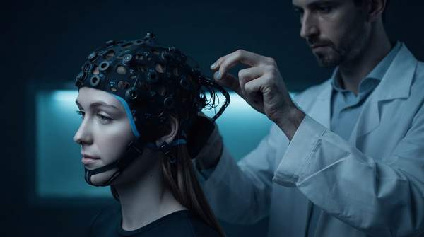
Choosing how a brain-computer interface acquires its signals is, at its core, a single decision about how much surgical risk a project can justify in exchange for neural data precision. Scalp EEG is non-invasive, cheap and safe, but its signals pass through skull and scalp, arriving noisy and spatially coarse. ECoG sits on the brain's surface under a craniotomy, capturing far cleaner signals at millimeter-scale resolution. The Utah Array penetrates cortical tissue to record individual neurons at roughly ten-micron resolution, powering the most demanding neuroprosthetics, but at the highest clinical risk. There is no best method—only the right point on that trade-off curve for a given project.
Understanding BCI Signal Acquisition: The Core Trade-offs
Researchers conventionally sort BCI signal acquisition methods by whether surgery is required, classifying them as non-invasive or invasive; a 2020 refinement by He and colleagues added a semi-invasive middle category, which is where ECoG typically lands. A review of BCI technologies frames the field's central tension plainly: each acquisition technique embeds trade-offs that must be matched to a specific application or user group rather than ranked in the abstract.
One useful way to think about the spectrum is the distinction a medical-engineering crossover review draws between "global" BCIs, which sample broadly distributed activity, and "local" BCIs, which detect precisely localized signals. Local systems capture more accurate signals, but because they depend on a limited number of neurons, they may not fully reflect complex cognitive processes. The same review notes that for domains like affective computing, implanting minute electrodes could actually impair the fidelity that biometric applications need. In other words, more precision is not automatically better—it must match what the application is trying to decode.
Non-Invasive EEG: Accessibility and Broad Applications
EEG places electrodes on the scalp, so signal acquisition happens entirely outside the body with no surgical risk. That safety profile is why a comparison study of BCI acquisition techniques calls EEG "the simplest, safest and most economic option." The price is paid in signal quality: EEG signals have a low signal-to-noise ratio and are attenuated by the skull and scalp, making them susceptible to noise and limiting detection of high-frequency oscillations.
Spatially, EEG is the coarsest of the three methods. A cross-pathway technical and clinical trade-off analysis puts scalp EEG's spatial resolution at roughly 10 millimeters, with negligible clinical risk, low cost and commercial maturity. Because EEG captures the synchronized activity of large populations of neurons rather than localized circuits, it suits applications where the goal is a broad readout—general cognitive state monitoring, consumer-grade devices, and basic research—rather than fine-grained control of complex hardware.
Semi-Invasive ECoG: Bridging the Gap in Signal Quality
Electrocorticography requires a craniotomy to place electrode arrays inside or outside the dura mater—subdural or epidural ECoG, respectively. The electrodes sit on the brain's surface without penetrating tissue, which is why the method is classified as semi-invasive: real surgery, but no direct cortical penetration.
The signal payoff is substantial. A BCI review notes that one of ECoG's primary benefits is its ability to detect high-frequency oscillations and other neural signals that are often lost or attenuated in scalp EEG recordings, an enhancement crucial for applications requiring precise temporal and spatial resolution such as motor and sensory mapping, language localization, and monitoring brain dynamics during cognitive tasks. Compared to EEG, subdural ECoG arrays deliver much higher spatial and temporal resolution, though they do not reach individual neuronal action potentials. The trade-off analysis assigns ECoG grids a spatial resolution of roughly 1 millimeter, with moderate clinical risk and high relative cost.
ECoG's clinical footprint is often tied to existing neurosurgical procedures. Conventional "macro" ECoG arrays used in intraoperative monitoring are predominantly handmade, with electrode diameters of several millimeters and inter-contact distances on the order of a centimeter—constraints that newer high-density thin-film arrays are designed to overcome. Because ECoG is less susceptible to relative micromotion of electrodes in tissue than single-neuron recordings, it offers a stability advantage over penetrating approaches.
Invasive Utah Array: Precision for Advanced Neuroprosthetics
The Utah Array is the canonical penetrating microelectrode array: a rigid grid of needle-sharp electrodes implanted directly into cortical tissue. Its manufacturer, Blackrock Neurotech, describes it as FDA-cleared and specifies up to 96 electrodes per array and up to 1,024 channels per system, recording and stimulating individual neurons or groups of neurons with what the company's product page calls "minimal invasiveness and maximum selectivity."
That marketing framing deserves scrutiny. Within the context of neural recording design, the array may be minimal for what it does, but academic and research contexts consistently classify penetrating microelectrode arrays as highly invasive, because the electrodes directly penetrate brain tissue. The cross-pathway analysis assigns the Utah Array single-unit spike recording at roughly 10 micrometers of spatial resolution, alongside high clinical risk—specifically glial scarring—high cost and investigational maturity. A research page from Beth Israel Deaconess Medical Center's Joshua Aronson calls the FDA-cleared Utah Array "the gold-standard electrode for monitoring neural activity in the human brain."
That precision comes with a durability caveat. A review of intracortical microelectrode arrays notes that while MEAs such as the Utah Array can function for months to years post-implantation, spike signal quality often degrades due to the immune response—an issue recent flexible MEA designs aim to address. Projects choosing the Utah Array should therefore plan for possible long-term signal decline, not just implantation risk.
BCI Signal Acquisition Methods: A Comparative Decision Framework
The table below contrasts the three technologies across the four dimensions that drive most selection decisions. Reading down the columns reveals the pattern that defines the field: as spatial resolution tightens from roughly 10 millimeters to roughly 10 micrometers, clinical risk climbs from negligible to high.
| Dimension | EEG | ECoG | Utah Array |
|---|---|---|---|
| Invasiveness / surgical risk | Non-invasive; scalp electrodes, no surgery, negligible risk | Semi-invasive; craniotomy to place electrodes on the cortical surface (subdural or epidural), no tissue penetration; moderate risk | Highly invasive; rigid electrodes penetrate cortical tissue; high risk including glial scarring |
| Signal quality (SNR) | Low SNR; signals attenuated by skull and scalp, susceptible to noise | High SNR; detects high-frequency oscillations lost in EEG | Superior SNR per the manufacturer; records single-unit spikes and local field potentials, though spike quality can degrade with immune response |
| Spatial resolution | Low, roughly 10 mm; large neural populations | Moderate, roughly 1 mm; does not reach single-neuron action potentials | Very high, roughly 10 µm; individual neurons |
| Primary application domain | Consumer-grade devices, cognitive state monitoring, basic research | Clinical monitoring such as seizure mapping, motor and language mapping, advanced research, emerging neuroprosthetics | Demanding neuroprosthetics and decoding of motion, vision and speech at investigational maturity |
Two qualifications keep this framework honest. First, the resolution figures come from a cross-pathway trade-off analysis and are approximate; the sources support relative ordering—low, moderate, very high—more firmly than exact numbers. Second, the Utah Array's "minimal invasiveness" label is the manufacturer's characterization of its recording design, not a clinical classification, and should not be read as comparable to EEG's risk profile.
Matching Technology to Application: A Decision Guide
The characteristics in the table translate directly into application fit. If the project goal is broad state monitoring—a meditation aid, attention tracking, or a consumer device that must be donned and doffed daily—EEG's safety, low cost and commercial maturity make it the only realistic choice, and its coarse resolution is not a limitation for that task.
If the goal is decoding fine motor function, sensation or speech, signal quality and channel count become the binding constraints. As the Aronson research group at Beth Israel Deaconess puts it, "the ability to capture neural patterns associated with fine motor function, sensation, and speech is dependent on signal quality and high channel count." That requirement pushes projects toward ECoG or intracortical arrays. ECoG is particularly attractive when a patient is already undergoing neurosurgery, since the monitoring opportunity comes bundled with an existing clinical procedure, and its resistance to electrode micromotion supports stable long-duration recording.
The Utah Array earns its invasiveness only when the application genuinely needs single-neuron resolution—high-performance neuroprosthetics such as robotic arm control, or detailed studies of neural network function. A BCI review notes that intracortical arrays are widely used for decoding motion, vision and speech, recording both low-frequency and spike signals. Teams choosing it should weigh the immune-response-driven signal degradation against the unmatched spatial precision, and note that the technology remains investigational rather than commercial.
Choosing Your BCI Path: Balancing Risk and Reward
Researchers and engineers should select a BCI signal acquisition method by carefully weighing the project's requirements for signal fidelity and spatial resolution against the acceptable level of surgical invasiveness and associated risks. Concretely: if the project needs only population-level readouts, EEG's negligible risk and low cost dominate; if it needs high-frequency signals and millimeter-scale mapping, ECoG's moderate risk buys a large quality gain; if it needs single-neuron decoding for a demanding neuroprosthetic, the Utah Array's high risk—including glial scarring and long-term spike degradation—is the price of roughly 10-micron resolution.
The measurable indicator of a sound choice is the project's success in achieving its BCI control or monitoring objectives, directly correlating with the chosen method's signal characteristics and the project's tolerance for risk. A team building a consumer attention monitor should watch whether EEG signal quality supports reliable state classification; a team decoding speech with ECoG should track whether high-frequency features remain decodable over recording sessions; a Utah Array project should monitor spike yield over months as the immune response unfolds. Matching the method to that objective at the outset—not after implantation or deployment—is what the trade-off curve rewards.
Sources
- A Review of Brain-Computer Interface Technologies: Signal Acquisition Methods and Interaction Paradigms — ARXIV
- Signal acquisition of brain–computer interfaces: A medical-engineering crossover perspective review - PMC
- BCI Systems and Comparison of Various Signal Acquisition Techniques — Multiresearchjournal
- Sensory Restoration via Brain-Computer Interfaces: A Unified 2×2 Framework and Convergence Roadmap — ARXIV
- Utah Array, Electrodes | Products — Blackrock Neurotech