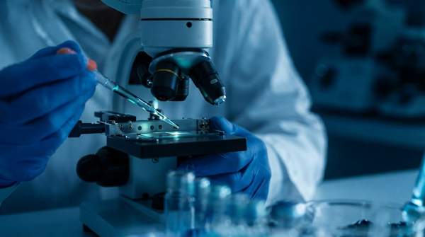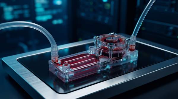
Organ-on-a-chip technology compresses living human tissue, blood-vessel-like flow, and mechanical forces into a device the size of a computer thumb drive. By culturing organ-specific cells inside microfluidic channels under continuous fluid flow, these systems can recapitulate organ-level physiology and disease with high fidelity—something neither flat petri-dish cultures nor animal models reliably achieve. The payoff for drug discovery is a preclinical model that predicts human therapeutic responses more accurately, while sharply reducing reliance on animal testing.
Introduction to Organ-on-a-Chip Technology
Donald E. Ingber, founding director of the Wyss Institute and author of a major review in Nature Reviews Genetics, frames the field by its central problem: "The failure of animal models to predict therapeutic responses in humans is a major problem that also brings into question their use for basic research." His answer is the organ chip—microfluidic devices lined with living cells cultured under fluid flow, which "can recapitulate organ-level physiology and pathophysiology with high fidelity."
The Wyss Institute, where much of the technology originated, describes the practical stakes: drugs that look safe and effective in animals frequently prove harmful or ineffective in humans, allowing useless or toxic compounds to advance through clinical trials at great expense while promising candidates never reach the market. Organ chips respond to that mismatch by modeling human biology directly, in vitro. A review of the technology's trajectory in the journal literature traces a surge of global interest to 2011, when a US presidential initiative on "human on chip" research was announced, drawing on cell biology, engineering, and materials science to simulate in-vivo tissues.
Core Components: Microfluidics and 3D Cell Cultures
The device itself is deceptively simple in concept. Emulate, a company spun out of the Wyss Institute, describes chips as flexible polymers containing parallel upper and lower microfluidic channels, each seeded with organ-specific cells. A thin, porous membrane separates the channels, creating an interface for cell-to-cell communication, and is coated with a tissue-specific extracellular matrix that helps drive tissue maturation. The whole assembly sits in a microfluidic platform that automates fluid flow and cyclic mechanical strain, maintaining physiologically relevant conditions in the chip's microenvironment.
Fluid flow does two jobs that static cultures cannot. First, it delivers nutrients and removes waste the way circulation does, sustaining metabolically demanding primary cells—a particular advantage for liver epithelial cells critical to toxicology studies, according to a review of three-dimensional culture systems. Second, moving fluid generates shear stress and, paired with the chip's flexible walls, cyclic mechanical strain that mimics the physical cues organs experience: the stretch of breathing lungs, the flexing of a beating heart wall. A PMC review of the technology notes that these systems offer accurate control of concentration gradients, fluid shear stress, tissue–tissue interfaces, and organ–organ interaction.
The cell culture side has moved beyond flat monolayers. A review in Frontiers in Bioengineering and Biotechnology explains that, in contrast to typical two-dimensional cultures, organ-on-chip platforms support 3D tissue constructs—cell aggregates, spheroids, and organoids—that can be formed first under standard protocols and then embedded in a gel-based matrix inside the chip's culture chambers. Earlier work showed how microfabrication borrowed from the microchip industry, combined with microfluidics, creates microenvironments that support tissue differentiation and recapitulate the tissue–tissue interfaces, spatiotemporal chemical gradients, and mechanical microenvironments of living organs. Induced pluripotent stem cells, introduced in 2006, became a common source of human cells for brain, heart, spinal cord, and kidney tissues.